
What are dental X-rays?
Radiography is an imaging technique that uses X-ray radiation to generate images of tissues and structures inside the body. An X-ray is a type of energy that passes through soft tissue and is absorbed by dense material. Therefore the X-rays pass easily through gums and cheeks (soft tissue) and are absorbed by teeth and bone (dense material). On X-ray images, dense materials show up as white, and soft tissue shows up as shades of grey.
Radiographs are an essential diagnostic and preventative tool that allow us to see inside the teeth, between the teeth, and below the gum line into the surrounding bone.
At VC Dental all our radiography imaging is low-radiation, digital and instant.
Why do I need X-rays?
There’s a lot going on inside your mouth that we can’t see from just a visual examination. During a visual examination we are only able to see the top part of your teeth that sits above the gum line. Even then, we often can’t see clearly between each of your teeth.
Radiographs allow us to see what’s happening between your teeth and below the surface, so we can perform a comprehensive ‘whole picture’ assessment of your oral health. This assists us in being proactive, rather than reactive, if there are any problems.
We utilise radiographs to perform many aspects of preventative and restorative dentistry, including: ruling out disease, diagnosing specific problems, evaluating the extent of a condition (such as dental decay), and planning the direction of treatment (such as wisdom tooth extraction).
Are dental X-rays safe?
The ADA (Australian Dental Association) has published that “Regardless of whether you’re a child or an adult, you can have X-rays safely taken of the inside and outside of your mouth. The amount of radiation involved is extremely low, and is equivalent to the sort of exposure you’d receive on a 1-2 hour flight. This means that even if you’re pregnant you can have X-rays taken, although they are generally kept to a minimum during this period”.
Types of X-rays
X-rays are divided into two main categories, intraoral and extraoral images.
An intraoral X-ray is taken inside the mouth.
An extraoral X-ray is taken outside of the mouth.
Intraoral images include:
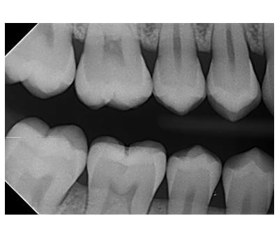
Bite wings
These are X-rays that are taken to detect interproximal (between adjoining tooth surfaces) cavities (decay).
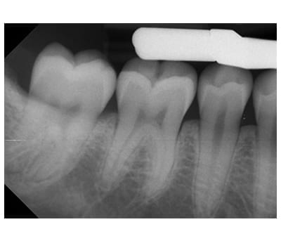
Periapical images
These are comprehensive X-rays taken of a specific tooth to identify localised problems. They whole the whole tooth from the crown (top) to beyond the end of the root to where the tooth is anchored in the jaw.
Extraoral images include:
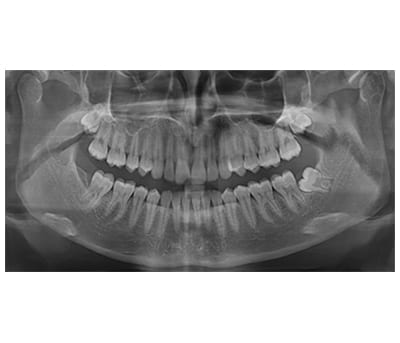
OPG
An OPG (Orthopantomogram) which is a two-dimensional wide panoramic image that displays all the teeth, the upper and lower jawbones and the sinus cavity in a single image. It shows the number, position and development of all the teeth including those that have not yet erupted. An OPG can be used in the planning of orthodontic treatment, dental implant surgery, wisdom teeth removal and root canal treatment. OPGs can also help diagnose cysts, jaw disorders and bone irregularities.
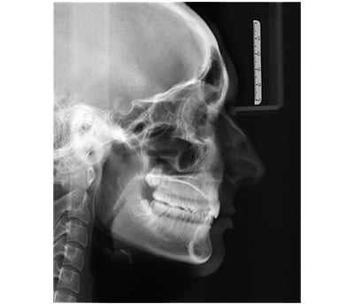
Lateral Cephalogram
A Lateral Cephalogram is a profile (side view) image of face. It includes your skull, soft tissues and facial contours in a single image and shows the relationship between your teeth and jaw bones. This image is usually taken at an orthodontic consultation (for example if you need braces).
What can X-rays detect?
Radiographs allow us to assess aspects of your teeth and mouth including:
- The health and density of your jawbone
- Your tooth roots and supporting tissues
- The condition of each tooth
- The condition of existing fillings/restorations
- The status of developing teeth
- The development of wisdom teeth
Through our assessment, we are able to diagnose conditions including:
- Presence and extent of tooth decay (which may otherwise be hidden between teeth)
- Problems with existing fillings, restorations, crowns or bridges
- Presence and extent of gum disease
- Presence and extent of bone loss
- Abnormalities in the soft tissue (such as abscesses or cysts)
- Some types of infections
- Whether developing teeth are present, and located in the correct areas
- Presence and location of wisdom teeth (for example if they are impacted)
- Traumatic injuries (for example tooth and/or bone fractures)
- The proximity of teeth to nerves and sinuses